New technique expands tissues so hundreds of biomolecules can be seen inside cells
Advertisement


For biologists, seeing is believing. But sometimes biologists have a hard time seeing.
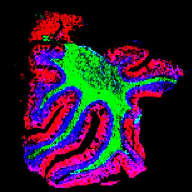
TEMI mapping of lipid distribution across cerebellar layers. This image shows lipid species specifically enriched in three distinctive layers of the cerebellum: molecular layer, white matter layer and granular cell layer.
Zhang and Ding et al.
One particularly vexing challenge is seeing all the molecules in an intact tissue sample, down to the level of single cells, simultaneously. Detecting the location of hundreds or thousands of biomolecules – from lipids to metabolites to proteins – in their native environment allows researchers to better understand their functions and interactions. Unfortunately, scientists don’t have great tools to accomplish this task.
Imaging methods, including most types of microscopy, provide a view of molecules inside cells. But they can track only a select handful of molecules at one time, and they can’t detect all types of biomolecules, including some lipids. Other methods, like regular mass spectrometry, can detect hundreds of molecules but don’t work on intact samples, so researchers can’t see how the biomolecules are oriented.

One promising technique – mass spectrometry imaging – overcomes some of these challenges. It allows researchers to see hundreds of molecules at one time in intact tissues. However, it doesn’t have high enough resolution to allow detection at the single cell level.
This was the problem Janelia Senior Group Leader Meng Wang faced. Wang and her team study the fundamental mechanisms behind aging and longevity, and they wanted to detect many different biomolecules in intact tissues to understand how the components change as tissues age.
“Knowing at each specific location what molecules are there and what is in the neighboring cells is very important for any kind of biological question,” Wang says.
Luckily, Wang’s lab is down the hall from Janelia Principal Scientist Paul Tillberg. Tillberg co-invented a technique called expansion microscopy as a graduate student at MIT. The method uses a swellable hydrogel material to expand samples uniformly in all directions to a point where fine details, like sub-organelle structure, can be detected with a conventional microscope.
Now a decade old, the expansion process is being applied to other methods outside traditional microscopy. Wang, Tillberg, and their collaborators at Janelia and the University of Wisconsin-Madison wanted to see if they could use expansion to overcome mass spectrometry imaging’s spatial resolution problem.
The result is a new method that expands tissue samples gradually without having to degrade them at the molecular level, as happens in the original expansion process. By expanding the intact samples in all directions, researchers can use mass spectrometry imaging to simultaneously detect hundreds of molecules at the single cell level in their native locations.
“This lets you have an untargeted look in the molecular space, and we are trying to bring it closer to what microscopy can do in terms of spatial resolution,” Tillberg says.
The team used the new technique to delineate the specific spatial patterns of small molecules in different layers of the cerebellum. They found that these molecules – including lipids, peptides, proteins, metabolites, and glycans – are not uniformly distributed, as previously thought. Moreover, they found that each specific layer of the cerebellum has its own signature of lipids, metabolites, and proteins.
The team was also able to detect biomolecules in kidney, pancreas, and tumor tissues, demonstrating that the method can be adapted for many different tissue types. In tumor tissues, they were able to visualize large variations in biomolecules, which could be useful for understanding the molecular mechanisms of tumors and potentially aid in drug development.
“When you can see these biomolecules, then you can start to understand why they have such patterns and how that is related to function,” says Wang. She believes the new technology will allow researchers to track these patterns during development, aging, and disease to understand how different molecules contribute to these processes.
Because the new method doesn’t require adding hardware to an existing mass spec imaging system, and the expansion technique is relatively easy to learn, the team hopes it will be used by many labs around the world. They also hope the new technique will make mass spec imaging a more useful tool for biologists and have laid out a detailed description of the new method and a roadmap for adapting it to other tissue types.
“We wanted to develop something that did not require specialized instruments or procedures, but can be broadly adopted,” Wang says.
Original publication
Other news from the department science


Most read news


More news from our other portals


Last viewed contents
What the sea spider genome reveals about their bizarre anatomy - The first high-quality pycnogonid genome provides novel insights in chelicerate evo-devo
More than 100-year-old flu virus sequenced - Swiss Genome of the 1918 Influenza Virus Reconstructed
Attracting bright and bold minds: new ideas for genome diagnostics and single molecule sensing - Four innovative life sciences ideas with particular economic potential receive awards
Deepest look yet into the human genome - A new resource for genome research worldwide

Researchers identify a potential biomarker for long COVID - Extracellular vesicles in study participants contain SARS-CoV-2 peptides

Qanatpharma, Zuse Institute Berlin, Enamine, and Proteros biostructures Announce Generative-AI Driven Lead Discovery Collaboration - Joint research program targets new treatments for life-threatening complication of brain hemorrhage

Merck (MSD) to acquire Verona Pharma for $10 billion - The acquisition strengthens the company's position in the field of COPD treatments

Takeda strengthens Berlin site as future German headquarters - The Constance site will be closed by the end of 2028
QIAGEN acquires bioinformatics company - Expanding clinical bioinformatics capabilities in molecular oncology

Remdesivir disrupts COVID-19 virus better than other similar drugs - How the COVID-19 drug remdesivir works at the molecular level

Scientists identify 160 new drugs that could be repurposed against COVID-19 - Forty of these drugs have already entered clinical trials